
Considerations When Planning Spatial Transcriptomics Analysis
The gold standard for gene expression profiling is bulk RNA sequencing (RNA-seq). RNA-seq performed on biologic samples reveals the specific genes that are differentially expressed under certain conditions. This provides a deeper understanding of the effects of different diseases, chemicals (drugs), or environmental factors. To complement the information obtained from bulk RNA-seq, researchers have developed single-cell RNA sequencing (scRNA-seq) techniques. As the name implies, scRNA-seq focuses on obtaining the RNA profiles of a specific type of cells extracted from a heterogeneous sample. This technique is used to study separate cell populations within a tissue or a tumor sample and/or to understand cell population dynamics. A similar technique for investigation into defined cell populations is Spatial Gene Expression Profiling (spatial GEP). As with scRNA-seq, there are several techniques, but broad commonalities in how the data are used.
What is Spatial Gene Expression Profiling?
Spatial Gene Expression Profiling investigates the RNA profiles of tissue subsections or cells taking into consideration their physical location within a larger tissue or biopsy sample. Researchers use spatial GEP techniques to gain insight into cellular biology and cell-to-cell dynamics based on gene expression within a two-dimensional or three-dimensional cellular microenvironment. Understanding a cell’s social and spatial context can be critical when the study goal is to capture the profile of cells in a specific region or exhibiting a certain characteristic.
How does Spatial Gene Expression Profiling work?
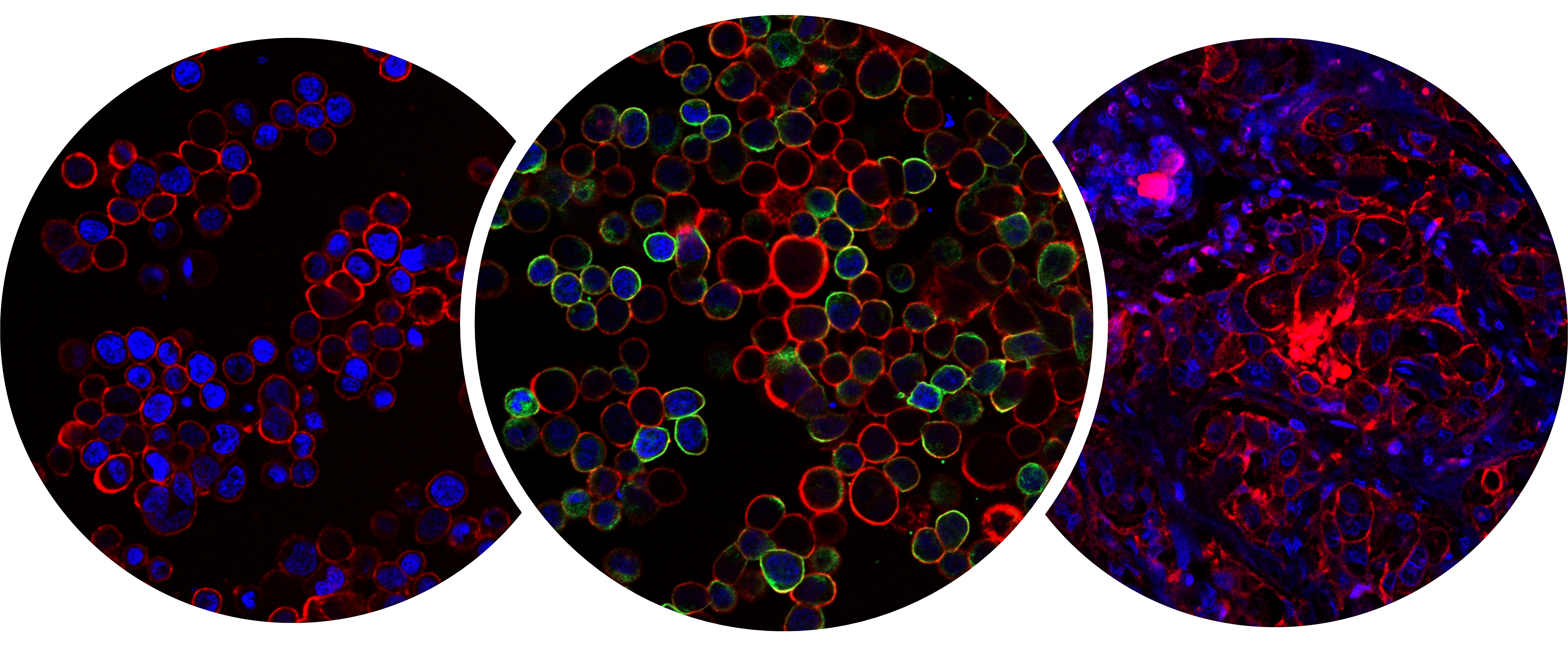
To conduct a spatial GEP experiment, scientists use a two-tiered approach combining immunofluorescence and gene sequencing. First, researchers map out the spatial gene expression using fluorescently labeled antibodies or probes, which bind to target proteins or RNAs. This allows researchers to identify their region of interest within a biopsy sample. The region of interest is exposed to ultra-violet light, causing the bound tags to illuminate. Finally, targets within the regions of interest, as identified by the tags, are collected and sequenced using next-generation sequencing, and an area-specific gene expression profile is thus obtained.
How does spatial GEP complement bulk RNA-seq?
In a recent Nature publication by Wilde D.C., et al. (2022) researchers used bulk RNA sequencing data from oropharyngeal squamous cell carcinoma (OPSCC) patients to train and validate a new Artificial Intelligence(AI) algorithm. The AI scoring system was intended to aid in risk assessment and development of precision oncology approaches for OPSCC patients in the future. In the same article the authors discuss the need for spatial GEP to complement the bulk RNA-seq testing. They discuss that combining bulk transcriptomic measurements with spatial resolution can reduce some of the inherent limitations of measuring gene expression across multiple cell types present in the same solid tumor. The study demonstrates that looking at data from complementing technologies can provide a much wider perspective to researchers, as well as serve to help confirm the findings across the two methods.
In another publication by Pachynski R.K., et al. (2021) the researchers state that about 15% of clinically significant prostate cancer (scPCa) cases cannot be visualized though a typical MRI. Their goal was to understand why some prostate cancers are invisible to MRI and identify methods for early diagnostics of these “invisible” scPCas. They used an oncology biomarker assay to conduct ultra-efficient bulk RNA sequencing of both normal and malignant tissue samples. Using these data, the group generated a gene expression profile of the relevant biomarkers and used these biomarkers as predictors of the disease. To complement and further advance the study, the researchers used single-cell spatial imaging and gene expression profiling in conjunction with AI-based analytic algorithms to examine the tumor ecosystem. They concluded that the stromal signature of the MRI-invisible scPCas were highly similar to that of the normal prostate tissue signatures, which is likely a contributing factor to the MRI invisibility. This study is the perfect example of how two complementing technologies can work hand in hand to provide answers to complex biological questions in a more efficient manner.
Summary
With new RNA sequencing technologies emerging, it is important to keep a broad perspective on how various methods can complement each other to expand the frontiers of transcriptomic research and precision medicine. While spatial GEP enables unprecedented specificity into certain cell types, it can also exclude valuable information contained in surrounding cells. Therefore, taking a multiprong approach to RNA sequencing may be the best path forward for many research initiatives. Obtaining data from bulk RNA sequencing, single-cell RNA sequencing, and spatial transcriptomics can provide a comprehensive image of not only the RNA profile of the tissue sample but also the cell-to-cell dynamics in a three-dimensional space. This model provides the closest layout for mimicking in vivo environment in vitro.